Bone Anatomy Lower Body / Bones Of The Lower Limb Teachmeanatomy - At the same time, the bones and joints of the leg and foot must be strong enough to support the body's weight while remaining.
Bone Anatomy Lower Body / Bones Of The Lower Limb Teachmeanatomy - At the same time, the bones and joints of the leg and foot must be strong enough to support the body's weight while remaining.. They also play a role in supporting the body's sexual functions, among other things. Inside the diaphysis is the medullary cavity, which is filled with yellow bone marrow in an adult. The lower leg contains two major long bones, the tibia and the fibula, which are both very strong skeletal structures. L1, l2, l3, l4, and l5. These bones are connected at the back with specialized joints.
The skeletal system also provides attachment points for muscles to allow movements at the joints. The femur is the single bone of the thigh. Fibrous tissue connects the lesser cornua to the body of the hyoid bone. Key bones in the abdominal area include the base of the ribcage and the lumbar spine in the lower back. A long bone has two main regions:
On This Page Muscles Of The Lower Limbnerves Of The Lower Limbjoints Of The Lower Limb There Are 64 Bones Anatomy Bones Medical Anatomy Skeletal System Anatomy from i.pinimg.com The lumbar spine connects to the thoracic spine above and the hips below. The femur is the single bone of the thigh. The skeleton acts as a scaffold by providing support and protection for the soft tissues that make up the rest of the body. It consists of 5 lumbar vertebra that are numbered 1 through 5 from top to bottom i.e. The lumbar vertebrae consist of five individual cylindrical bones that form the spine in the lower back. These are the femur, patella, tibia, fibula, tarsal bones, metatarsal bones, and phalanges (see link). The mandible (or lower jaw bone, latin: They also protect the delicate spinal cord and nerves within their vertebral canal.
The femur and/or hip may fracture secondary to trauma, so understanding the femur bone anatomy is important.
Distal to the ankle is the foot. Schau dir angebote von anatomie auf ebay an. Also known as the lower jawbone, the mandible is the largest and strongest bone of the face. The lumbar vertebrae consist of five individual cylindrical bones that form the spine in the lower back. The femur is the single bone of the thigh. At the same time, the bones and joints of the leg and foot must be strong enough to support the body's weight while remaining. The muscles of the lower back help stabilize, rotate, flex, and extend the spinal column, which is a bony tower of 24 vertebrae that gives the body structure and houses the spinal cord. A long bone has two main regions: Bones, discs, and joints in your lower back your lower back contains 5 vertebral bones stacked above each other with intervertebral discs in between. These vertebrae carry all of the upper body's weight while providing flexibility and movement to the trunk region. Mandibula) is the only movable cranial bone. The femur and/or hip may fracture secondary to trauma, so understanding the femur bone anatomy is important. The l5 vertebra is connected to the top of the sacrum (named the s1 segment) through an intervertebral disc.
They build the connection between the lower leg and the metatarsus. Each hip bone has three parts (ilium, ischium, pubis) and accepts the head of the femur to form the hip joint. Another essential function is to support the lumbar spine. The mandible (or lower jaw bone, latin: Lying exposed between the protective bones of the superiorly located ribs and the inferiorly located pelvic girdle, the muscles of this region play a critical role in protecting the.
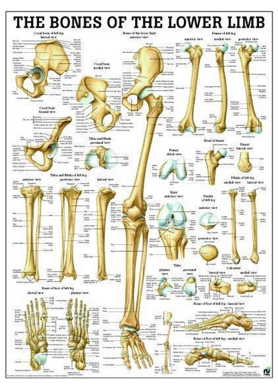
Bones Of Lower Limb Laminated Anatomy Chart from cdn11.bigcommerce.com It forms the foundation of the lower back and the pelvis. The tibia (also called the shinbone) is located near the midline of the leg. It consists of 5 lumbar vertebra that are numbered 1 through 5 from top to bottom i.e. Inside the diaphysis is the medullary cavity, which is filled with yellow bone marrow in an adult. Articulating at the knee and ankle joints respectively. The sacrum is a concave sphenoid bone that sits at the bottom of the spinal column. Anatomy of the pelvis and hip bones if you want to learn more about the hip joint and pelvic girdle, take a look below: The muscles of the abdomen, lower back, and pelvis are separated from those of the chest by the muscular wall of the diaphragm, the critical breathing muscle.
These vertebrae carry all of the upper body's weight while providing flexibility and movement to the trunk region.
The anatomy of the femur can be divided into proximal, central, distal, and posterior parts. Lying exposed between the protective bones of the superiorly located ribs and the inferiorly located pelvic girdle, the muscles of this region play a critical role in protecting the. They build the connection between the lower leg and the metatarsus. Key bones in the abdominal area include the base of the ribcage and the lumbar spine in the lower back. These muscles work together to produce movements such as standing, walking, running, and jumping. The tibia is the main bone of the lower leg, forming what is more commonly known as the shin. These are the femur, patella, tibia, fibula, tarsal bones, metatarsal bones, and phalanges (see link). The femur is a type of long bone located in the thigh and is the largest bone of the skeletal system. Glute max is one of the largest and most powerful muscles in the body. Each hip bone has three parts (ilium, ischium, pubis) and accepts the head of the femur to form the hip joint. Mandibula) is the only movable cranial bone. The sternum, or breastbone, is a flat bone at the front center of the chest. The lower limb contains 30 bones.
The anatomy of the femur can be divided into proximal, central, distal, and posterior parts. This article looks at the anatomy of the back, including bones, muscles. Key bones in the abdominal area include the base of the ribcage and the lumbar spine in the lower back. Inside the diaphysis is the medullary cavity, which is filled with yellow bone marrow in an adult. The sacrum is a concave sphenoid bone that sits at the bottom of the spinal column.
Lower Limb Bones The Human Skeletal System from sites.google.com The lower limb contains 30 bones. They also protect the delicate spinal cord and nerves within their vertebral canal. Bones also house bone marrow, which helps to produce a number of blood cell types that are vital to healthy body function. The anatomy of the femur can be divided into proximal, central, distal, and posterior parts. L1, l2, l3, l4, and l5. The femur is the single bone of the thigh. These are the femur, patella, tibia, fibula, tarsal bones, metatarsal bones, and phalanges (see link). The lower limb contains 30 bones.
These bones work together to provide flexibility to the trunk, support the muscles of the trunk, and protect the spinal cord and spinal nerves of the back.
The sacroiliac (si) joints connect the sacrum at the base of the spine with the hip bone. L1, l2, l3, l4, and l5. The l5 vertebra is connected to the top of the sacrum (named the s1 segment) through an intervertebral disc. The lumbar vertebrae consist of five individual cylindrical bones that form the spine in the lower back. Mandibula) is the only movable cranial bone. The muscles of the abdomen, lower back, and pelvis are separated from those of the chest by the muscular wall of the diaphragm, the critical breathing muscle. Inside the diaphysis is the medullary cavity, which is filled with yellow bone marrow in an adult. Distal to the ankle is the foot. They also play a role in supporting the body's sexual functions, among other things. Lying exposed between the protective bones of the superiorly located ribs and the inferiorly located pelvic girdle, the muscles of this region play a critical role in protecting the. The lumbar spine connects to the thoracic spine above and the hips below. The mandible (or lower jaw bone, latin: It consists of 5 lumbar vertebra that are numbered 1 through 5 from top to bottom i.e.
Another essential function is to support the lumbar spine anatomy lower body. These vertebrae carry all of the upper body's weight while providing flexibility and movement to the trunk region.


0 Komentar